

Electron Microscopy - Cell Culture and Tissue Samples
Ultra-thin section electron microscopy analysis is a specialized method whereby investigators can, with high resolution, directly observe the ultrastructure and morphology of cells, viruses, and tissues. This service allows investigators an opportunity to directly observe the minute, morphological details of particulate specimens, with unmatched clarity, resolution, and detail. It is unparalleled by any other technical method available today.
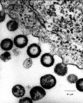 The ultrastructural details and morphology of specimens can be visualized and recorded by high-magnification photography, including intracellular and extracellular areas. Specimens are examined in the transmission electron microscope for ultrastructural characteristics, specifically to elicit the information required by the investigator, including;
- Ultrastructural characteristics of virus particles
- Viability and structural data on cells and tissues
- Condition and morphology of intracellular organelles
- Analysis of cell membranes, cell walls, and viral envelopes
- Examination for possible contaminants
- Morphological changes on cells over time
- Analysis on the effectiveness on virus-related vaccine and drugs
Numerous types of cells and tissues can be examined using this technique, including:
- Virus-infected cells
- Production cell lines
- Bacterial cells
- Fungi cells
- Yeast cells
- Biopsy or tissue samples
Service includes:
Service includes: Sample preparation using client-supplied cell supernatants. Each sample will be prepared multiple times, using various stains and sample dilutions. At least two grids per sample will be thoroughly examined in the electron microscope, to gain pertinent information based on our client’s requirements.
Final results include six to ten representative electron micrographs, and a final report documenting sample observations, analysis of the results, and materials and methods. Included is a CD containing the digital images of the samples, as well as the original negatives.
This email address is being protected from spambots. You need JavaScript enabled to view it.
See Electron Microscopy - Samples in Suspension for demo sample report